
A cavity may look like a chalky white patch, a brown or black mark, or a visible hole in a tooth. Early tooth decay may cause no pain, and cavities between teeth can be difficult to see. Not every mark indicates decay, so a dentist needs to examine suspicious changes.
Tooth decay begins when plaque bacteria produce acids that remove minerals from enamel. Early damage may leave the surface intact, but continued mineral loss can create a hole that needs treatment. At Enamel & Co., we focus on helping people understand daily oral care and enamel health. This guide explains how to recognize early cavity signs, identify hidden tooth decay, understand treatment options, and prevent future cavities.
What Do Cavities Look Like at Different Stages?
Cavities may look different as decay progresses: early mineral loss can appear as a chalky white patch, while later damage may leave a pit, hole, or broken tooth surface. There is no fixed color sequence. A dentist checks the condition of the enamel, not color alone.
According to the National Institute of Dental and Craniofacial Research (NIDCR), acids from bacteria remove minerals from enamel. The earliest changes may happen before a cavity, which is an actual hole, develops. Treating early mineral loss and repairing broken tooth structure are different tasks.
| Stage | What you might see | What is happening | What a dentist may recommend |
| Early mineral loss | Dull, chalky white patch | Enamel has lost minerals but its surface may remain intact | Risk assessment and preventive care |
| Surface breakdown | Small pit, rough area, or hole | The enamel surface has broken | Assessment for restoration or other care |
| Deeper decay | Larger opening or damaged edge | Decay may have reached dentin | Filling or a more extensive restoration |
| Advanced damage | Large broken area, sometimes without an obvious outer hole | Decay may be near or involve the pulp | Prompt assessment and treatment based on the findings |
Early Tooth Decay and Chalky White Spots
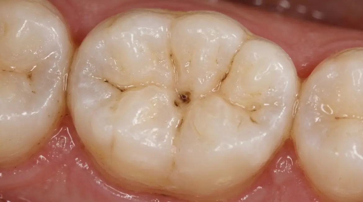
Early tooth decay may appear as a dull, chalky white spot that looks different from the shiny enamel around it. The surface may still feel smooth, and the tooth may not hurt. Bennett Amaechi studied early decay using human enamel samples in a laboratory. The early enamel lesion study showed how chalky white spots can develop after exposure to plaque bacteria.
Not every white patch indicates tooth decay. Dental fluorosis, enamel changes during tooth development, and older inactive lesions can cause similar marks. Early mineral loss may sometimes be stopped or reversed before a hole forms. However, laboratory findings cannot predict how quickly decay develops in a person’s mouth, so a dentist must check whether the area is active.
Small Cavities and Enamel Breakdown
A small cavity may look like a tiny pit, shallow hole, or damaged spot in the enamel. It can be hard to notice in a molar groove or between teeth, even under bright light. The size of the visible opening does not always show how far decay extends inside.
Once the surface breaks, bacteria and food can remain in the opening. A small cavity may not hurt, so waiting for a toothache is not a safe way to judge it. A dark line in a natural groove may also be a stain rather than a hole, which is why a dental examination matters.
Deep Cavities and Visible Tooth Damage
Deep decay may appear as a larger hole, a broken edge, or a dark damaged area, although the outside of the tooth can sometimes look less affected than the inside. Pain or sensitivity is possible when damage reaches deeper layers, but appearance and symptoms cannot measure the depth reliably.
Dentin lies beneath enamel, and the pulp inside the tooth contains nerves and blood vessels. When decay approaches or enters these tissues, the tooth may become painful or infected. A dentist examines the tooth and, when useful, takes an X-ray to decide how much structure can be saved.
What Does a Cavity Look Like on Different Teeth?
Some cavities are easier to see than others. Marks on front teeth may be visible when smiling, while cavities on back molars can hide in deep chewing grooves. Cavities between teeth may be difficult to notice because they develop on surfaces facing another tooth.
Dentists examine teeth from different angles and check areas where plaque collects. A tooth may look normal in the mirror even when decay is developing on a surface you cannot see. Dental X-rays may also help detect hidden cavities that are difficult to find during a visual examination.
Cavities on Front Teeth
A cavity on a front tooth may show as a spot or damaged area near the gumline, along an edge, or where two teeth touch. Since these teeth are visible when you talk and smile, you may notice changes sooner. However, not every white or brown mark indicates active decay.
White spots can develop around braces when plaque collects near the brackets. In a study of 332 orthodontic patients, Joshua Chapman found that 36% developed new white spot lesions on the upper teeth examined. Cleaning carefully around brackets and checking for new chalky marks can help you notice early enamel changes.
Cavities on Molars and Back Teeth
A cavity on a molar can be difficult to spot because the chewing surface has narrow grooves. You might see a pit or unusual area, but dark lines in those grooves can also be ordinary staining. Back teeth are hard to inspect without a dental mirror and good lighting.
Food and plaque can collect in deep pits and fissures. Dentists check these surfaces carefully and may recommend sealants for suitable teeth to lower the chance of future decay. If food repeatedly gets stuck in one molar or a surface feels damaged, have it examined rather than digging at it yourself.
Cavities Between Teeth
A cavity between teeth may have no visible mark on the surface you can see. It develops where neighboring teeth touch, so even a clear mirror or phone photo may miss it. A dentist may recommend bitewing X-rays when the examination and your cavity risk suggest hidden decay.
Dentists call decay on these contact surfaces interproximal caries. Food trapping or sensitivity in one area can prompt an examination, but neither symptom confirms that decay is present. Exposed tooth roots can also develop decay near the gumline when gums have receded, especially in older adults.
How Can You Tell a Cavity From a Tooth Stain?
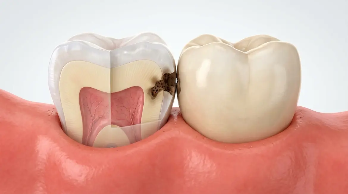
You cannot reliably tell a cavity from a stain by color alone. Coffee, tea, and other sources can darken enamel without creating a hole. Early decay may look white, while a brown or black mark might be staining or an inactive area of decay. A dentist checks the surface.
An outside stain can sit in a natural groove or cover several tooth surfaces. A cavity involves loss of tooth structure, although early mineral loss can happen before a hole develops. Look for changes worth reporting, such as a new mark, damaged edge, or area where food keeps catching, rather than trying to diagnose its color.
A white spot may also be dental fluorosis or a difference in enamel formation. An area of treated or arrested decay can stay dark even when it is no longer progressing. The dentist may clean and dry the tooth, inspect the surface, and use other tests if the diagnosis remains unclear.
How Do Dentists Find Cavities That Are Hard to See?
Dentists find hidden cavities by examining clean, dry teeth and checking hard-to-see surfaces. They may use bitewing X-rays or another approved imaging method when necessary. The decision depends on the patient’s symptoms, risk of decay, history, and what the dentist finds during the examination.
In a 2024 University of North Carolina laboratory, Adam Hoxie tested near-infrared imaging for early decay between extracted teeth. The interproximal imaging study found high specificity but only moderate sensitivity for that method. This means the scanner did not detect every early lesion in the study, so a single image cannot rule out decay.
What Does a Cavity Look Like on a Dental X-Ray?
On a dental X-ray, decay may show as a darker area within a tooth because damaged enamel or dentin allows more X-rays to pass through. A dentist studies the location and pattern of that shadow alongside the examination. A dark area alone is not a diagnosis.
Bitewing X-rays can reveal suspicious changes between the back teeth, where the contact surfaces are difficult to inspect. They may also help a dentist judge whether damage appears limited to enamel or extends toward dentin. Not every early lesion is visible, and image quality or overlapping tooth structures may limit what can be seen.
The American Dental Association (ADA) and the American Academy of Oral and Maxillofacial Radiology issued updated recommendations in January 2026. The guidance says dental imaging should follow a clinical examination and be ordered when it is needed for the individual patient. It does not call for the same X-ray schedule for everyone.
When Does Tooth Decay Need a Filling?
A filling is usually needed when decay has created a hole that cannot repair itself. Early mineral loss with an intact enamel surface may sometimes be stopped or reversed without drilling. A dentist decides after assessing the location, depth, and activity of the lesion. Saliva and fluoride can help replace minerals lost from early enamel decay. For an intact early lesion, a dentist may recommend changes in daily oral care, fluoride treatment, or another suitable preventive approach.
When a cavity needs repair, the dentist removes damaged material as appropriate and restores the tooth with a filling. A tooth with extensive damage may need a crown, while decay that reaches the pulp may require root canal treatment. In selected cases, dentists can use other methods to arrest decay. Not every dark spot needs drilling, and treatment must fit the tooth’s condition.
When Should You See a Dentist for Tooth Decay?
See a dentist if you notice a new hole, a damaged tooth surface, a persistent unusual mark, or repeated sensitivity. Early cavities may cause no pain, so do not wait for a toothache. A dental examination can confirm whether the change is tooth decay or another problem.
What does a cavity feel like? It may cause no symptoms at first, but later you may experience sensitivity to sweet, hot, or cold foods, pain when biting, or a lasting toothache. To understand how to know if you have a cavity, look for these changes and arrange an examination. Cracks and gum recession can cause similar symptoms.
Continue brushing gently and cleaning between your teeth, but avoid poking suspected holes or relying on phone photographs. Seek prompt dental care for severe pain, swelling, or fever. Trouble breathing or swallowing with facial or mouth swelling requires emergency medical attention because an infection may be spreading.
How Can You Prevent Tooth Decay in the Future?
You can lower your risk of tooth decay by brushing twice daily, cleaning between teeth, and reducing frequent sugary snacks and drinks. Fluoride toothpaste helps prevent cavities, while a dentist can recommend additional protection based on your age, tooth surfaces, and history of decay.
Plaque bacteria use sugars and starches to produce acids that remove minerals from enamelhttps://enamel-co.com/blog/can-toothpaste-strengthen-enamel-what-you-need-to-know/. Frequent sugar exposure increases these acid attacks, while saliva helps protect teeth. Preventive care can protect healthy tooth surfaces, but it cannot close an existing cavity.
Useful prevention habits include:
- Brush twice daily: Use a soft-bristled toothbrush and toothpaste suited to your dental needs.
- Clean between teeth: Use floss or a suitable interdental cleaner once a day.
- Limit sugary snacks: Reduce frequent sugary foods and drinks between meals.
- Ask about dental sealants: Sealants can help protect molars with deep chewing grooves.
- Keep dental appointments: Schedule examinations based on your dentist’s recommendations and cavity risk.
- Consider fluoride varnish for children: A dentist or pediatric medical professional may recommend it for children at higher risk of cavities. Read our guide to fluoride treatment for children to learn about its benefits and safety.
Support Healthy Teeth With Enamel & Co.
A good toothpaste supports everyday cleaning and enamel care, but it cannot fill a cavity. If you have a suspicious hole or active tooth decay, see a dentist rather than relying on toothpaste alone. Your choice of toothpaste should also reflect your personal cavity risk.
At Enamel & Co., we offer fluoride-free toothpaste with nano-hydroxyapatite in an airless pump. Our dentist-owned brand works with participating dental offices in Mesa, Gilbert, and San Tan Valley, Arizona. Nano-hydroxyapatite is studied for enamel remineralization, but that does not mean our toothpaste can repair an established cavity.
Ready to choose toothpaste for your daily routine? Shop fluoride-free toothpaste and review the current formula and buying options. Customers in Arizona and elsewhere should ask a dentist about their cavity risk, especially if they have ongoing decay or need extra cavity protection.
Conclusion
A cavity may begin as a dull white patch and later become a pit or hole, but not every dark or white mark is decayed. Some cavities are hidden between teeth, and early tooth decay may cause no pain. Color alone cannot confirm the problem.
If you notice a new change, have a dentist assess it before deciding what treatment is needed. Early mineral loss may sometimes be stopped, while an established hole generally needs repair. Daily cleaning and suitable preventive care can lower the risk of new cavities.
Frequently Asked Questions
Can a Small Hole in a Tooth Be Something Other Than a Cavity?
Yes. A small dip may be a natural groove, a chipped area, or an enamel defect rather than a cavity. A dentist can check whether tooth structure has been damaged by decay or another cause. Do not probe the opening with a sharp object.
Can Cavities Develop Around Old Fillings or Crowns?
Yes. Decay can develop in natural tooth structure beside a filling or crown, particularly near its edge. The restoration itself does not decay like enamel, but bacteria can affect the tooth around it. The area may be difficult to see without a dental examination.
Do Cavities Look Different in Baby Teeth?
Yes. Cavities in baby teeth can show as white areas, dark spots, or holes, just as they do in permanent teeth. Young children may develop decay on their upper front teeth and molars, and they may not complain of pain early on.
How Quickly Can a Small Cavity Get Worse?
There is no single number of days or weeks that predicts how quickly a small cavity will worsen. Progress depends on diet, saliva, plaque control, tooth location, and whether the lesion is active. A dentist can assess these factors and recommend follow-up or treatment.
Can You Check for Cavities Using a Phone Camera?
No. A phone camera may show a mark or visible hole, but it cannot reliably confirm a cavity. Shadows, lighting, and tooth position can change what appears in a photo. Decay between teeth may not show up at all.